WNK (With No Lysine [K]) kinases are serine/threonine kinases implicated in familial hyperkalemic hypertension (FHHt). Beyond blood pressure regulation, WNKs are emerging therapeutic targets for stroke and certain cancers, including triple-negative breast cancer and glioblastoma. In this study, we aimed to identify and characterize novel inhibitors of WNK kinases. A high-throughput screening of approximately 210,000 compounds was performed, followed by compound re-acquisition, secondary assays, commercial selectivity profiling, and crystallographic analysis to discover inhibitors with isoform-specific activity against WNK kinases. Five distinct classes of compounds were found to inhibit WNK1 kinase activity: quinoline derivatives, halo-sulfones, cyclopropane-containing thiazoles, piperazine-containing compounds, and nitrophenol-based molecules. These compounds broadly inhibit all four WNK isoforms, demonstrating strong pan-WNK selectivity. Notably, a subset of quinoline derivatives displayed isoform-selective potency, with greater inhibition of WNK3 compared to WNK1. Structural analysis of the quinoline compound SW120619 bound to WNK3’s kinase domain revealed active site engagement, and comparison with WNK1 provided insights into the molecular basis of isoform specificity. The identified compound classes provide promising scaffolds for the development of pharmacological tools and potential therapeutic agents targeting hypertension and cancer.
Introduction
WNK (With No Lysine [K]) kinases are cytoplasmic serine/threonine protein kinases distinguished by their unique arrangement of catalytic residues [1]. They play critical roles in transepithelial ion transport, regulation of cell volume, and cell motility [2–5], establishing them as promising therapeutic targets for multiple diseases. WNK1 and WNK4 are linked to familial hyperkalemic hypertension (FHHt), also known as Gordon Syndrome [6–8], while knockout or knockdown of WNK1 and WNK3 reduces blood pressure [9, 10], supporting their relevance for hypertension treatment. Additionally, WNK kinases are implicated in cancers including breast, lung, ovarian, and brain malignancies [11–14]. Notably, transposon-based studies identify WNK1 as a proto-oncogenic signature gene in triple-negative breast cancer [15], while WNK3 knockout mice display reduced edema in stroke models [16, 17] and show overexpression in the hippocampus in certain epileptic conditions [18].
Targeting WNK kinases with small-molecule inhibitors can elucidate their biological roles and serve as starting points for drug development. Previous efforts have yielded pan-WNK inhibitors such as WNK463, which is ATP-competitive [19, 20], and allosteric inhibitors like WNK476 that bind adjacent to helix C [21]. Here, we describe novel scaffolds that selectively inhibit WNK1 and WNK3, and crystallographic analysis of a WNK3-specific inhibitor provides insights into the structural basis of isoform selectivity.
Materials and Methods
Reagents
Kinase-Glo® luminescence reagent was purchased from Promega Inc. A plasmid encoding a GST-tagged OSR1 substrate peptide (GST-TEV-GAM314RAKKVRRVPGSSGRLHKTEDGGWEWSDDEF344) was kindly provided by Melanie Cobb (UT Southwestern Medical Center, unpublished) and validated by sequencing and tandem mass spectrometry. WNK1 inhibitors identified from the screen were obtained from ChemBridge, ChemDiv, ComGenex, Prestwick, Collaborative Drug Discovery, and UT Southwestern synthesis labs. Recombinant WNK1 (residues 1–491) and WNK3 (1–431) for mobility shift assays were obtained from Carna Biosciences USA. Fluorescein-labeled OSR1 peptide (FAM-RVPGSSGRLHK-NH2) was purchased from Bachem America.
Protein expression and purification
The pWNK1 kinase domain (residues 194–483) was expressed in E. coli following the protocol of Min et al. [22], with benzonase and protease inhibitors added during lysis. Approximately 800 mg of purified protein was obtained from 25 preparations, with phosphorylation at Ser382 confirmed by mass spectrometry. A codon-optimized pWNK3 (118–409) construct was expressed and purified using the same method, with Ser308 phosphorylation verified [23]. GST-OSR1 (314–344) peptide was expressed in Rosetta (DE3) pLysS E. coli and purified with glutathione beads, yielding 3 g of substrate for screening.
High-throughput screening
A total of 210,000 compounds were screened in 384-well plates using the Kinase-Glo® assay (Promega) at the UT Southwestern HTS Core. Each well contained substrate solution (55.3 mM HEPES pH 7.4, 55.3 mM MgCl2, 4.7 µM pWNK1, 19.3 µM GST-OSR1) with 0.2 µL DMSO (negative control) or 0.5 mM compound. ATP was added to a final concentration of 21 μM, plates were incubated for 2 h at room temperature, and luminescence was read using an EnVision plate reader (PerkinElmer).
Compound library
The UT Southwestern chemical library comprises ~210,000 compounds from multiple vendors, including ChemBridge (75,000), ChemDiv (100,000), ComGenex (22,000), TimTek (1,200), Prestwick (1,100), and the NIH Clinical Collection (450). An additional 2,500 compounds were synthesized in-house. Compounds largely comply with Lipinski’s rules (MW <550 Da), and the library contains ~6,500 partially purified natural product fractions from marine bacteria.
Data analysis and hit confirmation
Screening data were analyzed using Genedata Screener® v10.1. Top hits with Z-scores >3σ were rescreened for pWNK1 and pWNK3 at 6, 4, and 0.5 μM. IC50 values were further refined for the 96 most potent compounds using nine concentrations (50 nM–49.5 μM).
Mobility-Shift assays
Kinase activity was measured via phosphorylation-induced mobility shifts of FAM-OSR1 substrate peptides in 384-well plates. GST-WNK1 or GST-WNK3 (12.5 nM final) was pre-incubated with inhibitors (0.04–10 μM) before initiating reactions with 0.5 μM FAM-OSR1 peptide and 50 μM ATP. Reactions were incubated 3 h, quenched, and analyzed using a PerkinElmer LabChip® EZ reader.
Specificity screening
SW133708, SW120619, and SW182086 were evaluated against 50 kinases spanning all five classes by Eurofins Inc. at 10 μM compound concentration using MBP or native substrates.
Crystallography
WNK3-KDm/S308A was co-crystallized with SW120619. Crystals grew at 16 °C in 0.1 M Na Malonate pH 5.0, 12% PEG3350, and were cryoprotected in 20% glycerol. Diffraction data (3.1 Å) were collected at APS 19-ID. Structures were solved by molecular replacement (PDB 5DRB) and refined using REFMAC; ligand modeling was performed in Coot. Structure coordinates are deposited under PDB 8EDH.
Differential scanning fluorimetry
pWNK1 or pWNK3 (5 μM) was incubated with 0.5 mM inhibitor, 50 mM HEPES pH 7.5, 150 mM NaCl, and 5× SYPRO Orange in 25 μL wells. Temperature was ramped from 4–80 °C in 0.5 °C increments on a Bio-Rad CFX96, monitoring fluorescence in the fluorescein channel [24].
Results and Discussion
Bootstrap screens
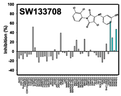
The phosphorylated WNK1 kinase domain (pWNK1-KDm) expressed in E. coli was employed for inhibitor screening. A bootstrap approach was implemented to identify WNK1 inhibitors within the UT Southwestern chemical library. At the time of the screen, the only reported WNK1 inhibitor, hypericin (US Patent Application US 2008/0286809 A1, Dario Alessi), exhibited an IC50 of 200 μM. To establish a more effective positive control, a focused sub-library of 3,500 representative compounds was screened using myelin basic protein as the substrate. This small-scale screen identified SW133708 as a promising hit. Subsequent commercial specificity testing (Eurofins) confirmed that SW133708 displayed potent pan-WNK activity, inhibiting WNK1, WNK2, and WNK4 [25].
SW133708 was then applied as a reference in a slightly larger 8,000-compound screen, which identified SW137446 as a major hit. SW137446 subsequently served as the positive control in the full 210,000-compound screen. Both the 8K and 210K screens utilized a GST-fusion construct containing the pan-WNK phosphorylation site of OSR1 [26, 27]. Performing the full screen required 3 g of the OSR1 fusion peptide and 800 mg of pWNK1-KDm.
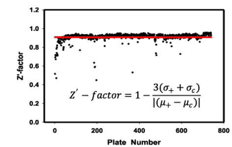
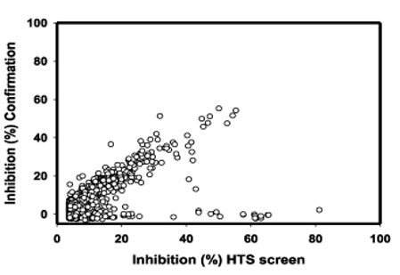
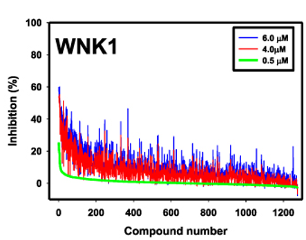
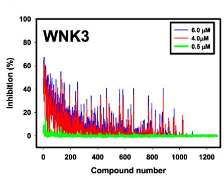
The Z′-factor for both the 8K and 210K screens was 0.91, indicating robust separation between positive and neutral controls across plates, with only seven plates falling below this threshold (Figure 1b). From the 210K screen, the top 1,275 compounds exhibiting Z-scores ≥3σ were re-screened in triplicate at concentrations of 6 μM, 4 μM, and 0.5 μM (Figures 1c and 1d). The Z′-factor for the 36 confirmation plates was 0.94, demonstrating excellent assay reliability. Results from the confirmation screen closely correlated with the initial HTS data, as evidenced by the diagonal pattern observed (Figure 1c). Dose-dependent inhibition was further confirmed (Figure 1d), reinforcing the consistency between the 210K primary screen and the confirmation screen.
Finally, 96 selected compounds were re-screened across nine concentrations to refine IC50 values against pWNK1. Representative data are presented in Figures 2–6.
|
|
|
|
|
|
a) |
b) |
c) |
|
|
|
|
||
|
d) |
e) |
||
|
Figure 1. High-throughput and specificity screen results. (a) Commercial specificity screen (Eurofins, Inc.) testing the cross-reactivity of SW133708, the initial hit from the 3,500-compound screen, against 50 diverse kinases. (b) Z′-factor for each plate of the 210,000-compound screen (blue) is shown, with the average Z′-factor across all plates indicated by the red line. (c) Scatter plot of % inhibition in the confirmation screen versus % inhibition in the original high-throughput screen, demonstrating consistency between screens. (d) % inhibition of each compound at 6 μM (blue), 4 μM (red), and 0.5 μM (green) toward WNK1. (e) % inhibition of each compound at the same concentrations toward WNK3. |
|||
|
|
|
Figure 2. Quinoline-derived inhibitors identified from the 210,000 compound screen. IC50 WNK1 and IC50 WNK3 are calculated using non-linear regression method in GraphPad from three inhibitor concentrations (6 μM, 4 μM and 0.5 μM). IC50WNK1* is calculated from the refined HTS screen conducted at nine inhibitor concentrations. |
|
|
|
Figure 3. Halo-sulfone inhibitors identified from the 210,000 compound screen. IC50 WNK1 and IC50 WNK3 are calculated from three inhibitor concentrations (6 μM, 4 μM and 0.5 μM). - indicates compounds that do not meet a 3 σ criterion for the confirmation screen |
|
|
|
Figure 4. Cyclopropane-containing thiazole inhibitors identified from the 210,000 compound screen. IC50 WNK1 and IC50 WNK3 are calculated from three inhibitor concentrations (6 μM, 4 μM and 0.5 μM). IC50 WNK1* is calculated from the refined HTS screen conducted at nine inhibitor concentrations. |
|
|
|
Figure 5. Nitrophenol-derived inhibitors identified from the 210,000 compound screen. IC50 WNK1 and IC50 WNK3 are calculated from three inhibitor concentrations (6 μM, 4 μM and 0.5 μM). IC50 WNK1* is calculated from the refined HTS screen conducted at nine inhibitor concentrations. |
|
|
|
Figure 6. Piperazine-derived inhibitors identified from the 210,000 compound screen. IC50 WNK1 and IC50 WNK3 are calculated from three inhibitor concentrations (6 μM, 4 μM and 0.5 μM). IC50. WNK1* is calculated from the refined HTS screen conducted at nine inhibitor concentrations |
Using the screening facility, the confirmation screen was cross-analyzed with phosphorylated kinase domain of WNK3 (pWNK3-KDm (pWNK3)). This screen gave similar results with respect to quality and correlation as the WNK1 screen.
Identification of compound classes
Through high-throughput screening, five distinct structural classes of WNK inhibitors were discovered: quinoline derivatives (Figure 2), halo-sulfones (Figure 3), cyclopropane-containing thiazoles (Figure 4), piperazine derivatives (Figure 5), and nitrophenol derivatives (Figure 6). Representative compounds from each class, along with their corresponding inhibitory potencies against pWNK1 and pWNK3, are presented in Figures 2–6. Notably, quinoline-derived inhibitors displayed a preference for WNK3 over WNK1, as detailed in subsequent analyses. The halo-sulfone class is described separately elsewhere [28]. The cyclopropane-thiazole series, as well as the piperazine and nitrophenol compounds, exhibited variable activity across WNK isoforms.
Selectivity assessment
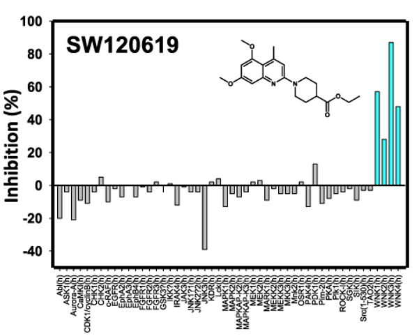
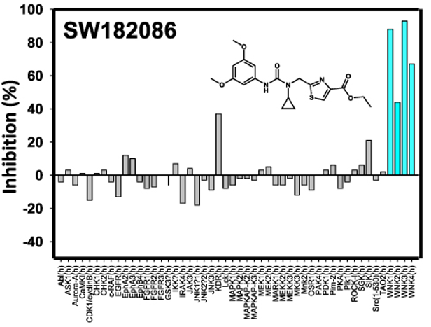
Two inhibitors, SW120619 (from the quinoline class) and SW182086 (from the cyclopropane-thiazole class), were further evaluated at 10 μM for kinase selectivity against a panel of 46 protein kinases and all four WNK isoforms (Eurofins Inc., France); (Figure 7). Both compounds demonstrated high specificity toward WNK kinases. Specifically, SW120619 preferentially inhibited WNK3, followed by WNK1, WNK4, and WNK2, whereas SW182086 showed comparable inhibitory activity against WNK1 and WNK3. These findings highlight the potential for developing isoform-selective WNK inhibitors.
|
|
|
|
a) |
b) |
|
Figure 7. Kinase selectivity assessment |
|
The kinase selectivity of the most potent compounds from the quinoline and cyclopropane-thiazole classes was evaluated using a commercial screen (Eurofins Inc.) at a fixed concentration of 10 μM. The four human WNK isoforms are indicated by blue lines in the figure.
Quinoline class inhibitor activity
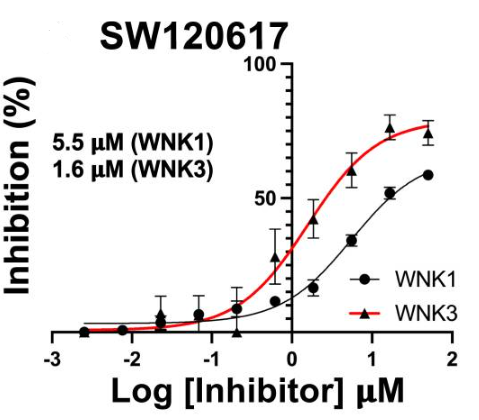
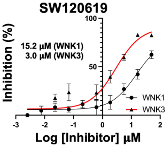
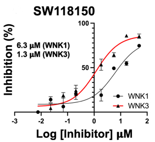
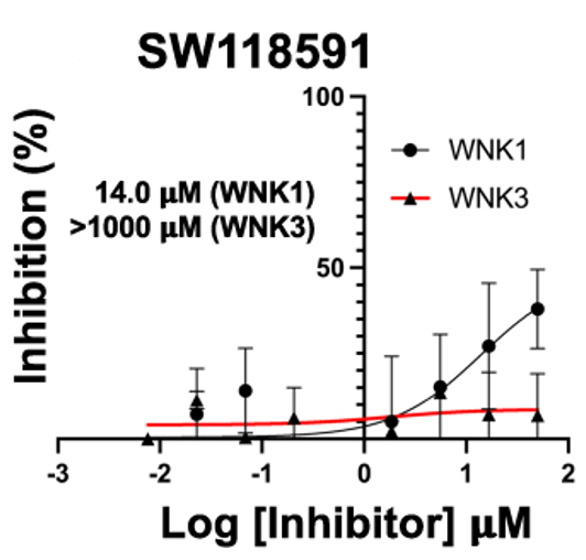
Four quinoline-derived compounds identified in the high-throughput screen were obtained from commercial vendors and tested using peptide mobility shift assays. Among these, SW120617, SW120619, and SW118150 exhibited stronger inhibition of WNK3 relative to WNK1 (Figures 8a–8c). In contrast, SW118591 displayed minimal or negligible inhibitory activity against WNK3 and WNK1 (Figure 8d), indicating variable potency within this chemical series.
|
|
|
|
a) |
b) |
|
|
|
|
c) |
d) |
|
Figure 8. Peptide mobility shift assays for quinoline class inhibitors. (a) SW120617, (b) SW120619, (c) SW118150, and (d) SW118591. GST-WNK1 (black line) and GST-WNK3 (red line) using a fluorescently labeled FAM-OXSR1 substrate peptide. |
|
Compound characterization and specificity analysis
High-throughput screening identified five principal chemical classes that inhibit WNK kinases: quinoline derivatives, halo-sulfones, cyclopropane-containing thiazoles, piperazine derivatives, and nitrophenol-based compounds. Representative compounds from each class were selected for detailed evaluation, with their chemical structures and inhibitory activity against pWNK1 and pWNK3 presented in Figures 2–6. Among these, the quinoline derivatives displayed preferential inhibition of WNK3 over WNK1. The halo-sulfone series is the subject of a separate study [28]. Other compound classes included cyclopropane-thiazoles, piperazine derivatives, and nitrophenol compounds.
To determine selectivity, two inhibitors — SW120619 (quinoline class) and SW182086 (cyclopropane-thiazole class) — were tested at 10 μM against a panel of 46 kinases along with the four human WNK isoforms (Figure 7). Both compounds demonstrated high selectivity toward WNK kinases. SW120619 showed a rank order of potency of WNK3 > WNK1 > WNK4 > WNK2, whereas SW182086 exhibited comparable activity against WNK1 and WNK3.
Four quinoline-class inhibitors were further examined using peptide mobility shift assays. SW120617, SW120619, and SW118150 inhibited WNK3 more strongly than WNK1 (Figures 8a–8c), while SW118591 had minimal or negligible inhibitory effects on both kinases (Figure 8d).
Differential scanning fluorimetry (DSF) was performed to assess binding-induced thermal stabilization. All four quinoline derivatives increased the melting temperature (Tm) of both pWNK1 and pWNK3, with ∆Tm values ranging from 0–2.5°C for pWNK1 and 6.5–14°C for pWNK3 (Table 1), indicating stronger binding to WNK3. These thermal shifts were consistent with the IC50 data from mobility shift assays, supporting the observed isoform selectivity of these compounds.
Table 1. Change in melting temperature Tm (∆Tm) upon binding of quinoline-derived compounds to pWNK1 and pWNK3 using differential scanning fluorimetry
|
Inhibitor |
∆Tm pWNK1+Inhibitor (°C) |
∆Tm pWNK3+Inhibitor (°C) |
|
SW120619 |
2.0 |
7.5 |
|
SW120617 |
1.5 |
11.0 |
|
SW118150 |
2.5 |
14.0 |
|
SW118591 |
0.0 |
6.5 |
Crystal structure of SW120619 bound to WNK3/SA
The primary autophosphorylation site of WNK3, Ser308, was mutated to alanine (S308A) to prevent autophosphorylation, generating WNK3/SA. The X-ray structure of the WNK3/SA kinase domain in complex with the quinoline-derived inhibitor SW120619 was determined at 3.1 Å resolution by molecular replacement using the WNK1/SA structure (PDB: 6CN9) as a search model.
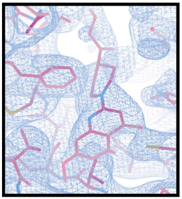
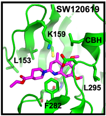
SW120619 (ethyl 1-(5,7-dimethoxy-4-methylquinolin-2-yl) piperidine-4-carboxylate) consists of a quinoline ring fused to a piperidine scaffold and is well-resolved in the electron density (Figure 9a). The inhibitor adopts an orientation roughly perpendicular to the β-strands of the N-terminal lobe, engaging two ridge-like regions of the β-sheet. On one ridge, SW120619 interacts with L153, V161, C176, and L225, while on the second ridge, it contacts A174 and T227. Additionally, it engages the backbone of residues L229–T231 located at the crossover connection between the N- and C-terminal lobes (Figure 9b).
On the opposite face of the kinase, SW120619 forms extensive interactions with F282 and establishes contacts with the catalytic residues K159 and D294. Its methoxy substituents interact with the chloride-binding helix (CBH) at the N-terminal region of the activation loop (previously described as the “3₁₀ helix”), specifically contacting the main chain of L295 and the side chain of L297 (Figure 9b).
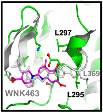
Structurally, SW120619 resembles the pan-WNK inhibitor WNK463 reported by Novartis, both in overall molecular architecture and binding orientation (Figure 9c). Like WNK463, SW120619 consists of a linear arrangement of heterocyclic rings and occupies the same binding pocket near the crossover connection. However, SW120619 does not extend as deeply into the active site as WNK463.
|
|
|
|
|
a) |
b) |
c) |
|
|
|
|
|
d) |
e) |
f) |
|
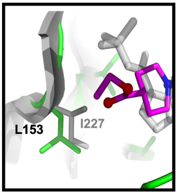
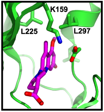
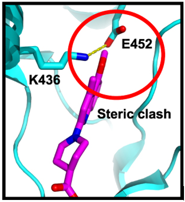
Figure 9. Structure of complex between WNK3/SA and SW120619. (a) Electron density of the inhibitor SW120619 contoured at 0.5 σ. (b) Schematic of the inhibitor binding drawn in Pymol (c) Superposition of WNK3/SW120619 with WNK1/6CN9. The superposition encompassed residues forming the binding site, WNK1/225–236, 298–308, 353-375 (in WNK3, WNK3/151-162, 223–233, 279-301) (d) Closeup of SW120619 and WNK463 near L153 in WNK3 (I227 in WNK1). (e) The WNK3/SW120619 complex oriented to highlight the differences between WNKs and more standard protein kinases such as PAK6 (PDB file 2C30). (f) PAK6 oriented in comparison with (e). Red circle indicates steric clash between SW120619 and PAK6. |
||
Structural basis for WNK3 selectivity of SW120619
Comparison of WNK3 and WNK1 residues contacting SW120619 revealed a single amino acid difference, with leucine at position 153 in WNK3 corresponding to isoleucine 227 in WNK1 (Figure 9d). This substitution, combined with the specific binding orientation of SW120619, likely contributes to the observed WNK3 selectivity. To further evaluate the binding mode, the WNK1 structure complexed with WNK463 was locally superimposed onto WNK3/SW120619 using secondary structure elements that define the inhibitor binding site (Figure 9c). The superposition shows that SW120619 makes direct contact with WNK3/L153, whereas WNK463 does not (Figure 9d). Additional subtle variations in the chloride-binding helix (CBH), including L295 and L297 in WNK3, may also underlie the selective binding of SW120619.
The enhanced potency of quinoline-derived inhibitors toward WNK3 suggests the feasibility of developing isoform-specific compounds. Analysis of the WNK3/SW120619 structure compared with canonical serine/threonine kinases, such as PAK6, indicates that the inhibitor occupies a pocket that is sterically incompatible with standard kinase structures (Figures 9e–9f). In WNK3, the pocket accommodating the quinoline moiety is accessible due to the unique positioning of residues and the absence of a steric clash seen in PAK6. These observations rationalize both the pan-WNK activity and the potential for isoform selectivity of quinoline-based inhibitors.
Crystallographic data indicate that SW120619 occupies the same pockets as the pan-WNK inhibitor WNK463 but engages a smaller subset of binding sites (Pockets I and II). Modeling of other quinoline analogues highlights potential positions for chemical modification, supporting rational design approaches to enhance isoform specificity. This structural insight provides a foundation for the development of selective WNK1 or WNK3 inhibitors.
Conclusion
Through high-throughput screening, we identified selective inhibitors of WNK1 and WNK3 with strong specificity toward WNK kinases over a panel of 46 other kinases. Structural comparisons between WNK isoforms and canonical serine/threonine kinases reveal active site differences that account for this selectivity. Quinolinederived compounds exhibited higher potency against WNK3 compared with WNK1, and structural analysis provides guidance for designing isoform-specific inhibitors. These inhibitors serve as valuable starting points for developing pharmacological tools to investigate isoform-specific WNK functions in signal transduction and their roles in hypertension, cancer, and other diseases.
Acknowledgments: We thank Clinton Taylor and Melanie Cobb for the gOSR1 peptide. We thank Anwu Zhou for help in analyzing the HTS data. Results shown in this report were derived from work performed at Argonne National Laboratory, Structural Biology Center (SBC) at the Advanced Photon Source. The SBC is operated by the U Chicago Argonne, LLC, for the US Department of Energy, Office of Biological and Environmental Research under contract DE-AC02-06CH11357. Crystallographic studies were coordinated by Diana Tomchick in the UT Southwestern Structural Biology Laboratory. We thank Kenneth Westover for helping with the mobility-shift assays.
Conflict of Interest: None
Financial Support: This work was supported by the American Heart Association grants 16SA285300002 and 14GRNT20500035, the Welch Foundation grants I1128 and I-2100-20220331, and CPRIT grant RP190421 to EJG and a pilot grant from Harold C. Simmons Comprehensive Cancer Center to EJG and RA.
Ethics Statement: None